Home
Uncategories
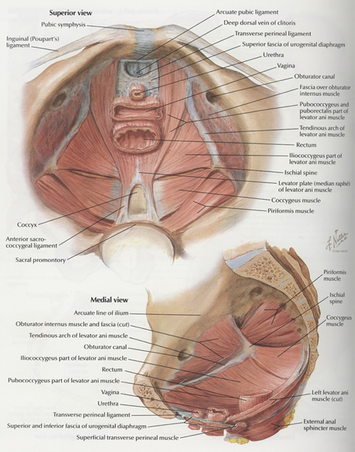
Anatomy Muscles Pelvis : Pelvic floor muscles as seen from below in supine female subject [taken... | Download Scientific ... / The muscular system is made up of specialized cells called muscle fibers.
Anatomy Muscles Pelvis : Pelvic floor muscles as seen from below in supine female subject [taken... | Download Scientific ... / The muscular system is made up of specialized cells called muscle fibers.
Anatomy Muscles Pelvis : Pelvic floor muscles as seen from below in supine female subject [taken... | Download Scientific ... / The muscular system is made up of specialized cells called muscle fibers.. (1) the obturator internus and the the fascia of the obturator internus covers the pelvic surface of, and is attached around the margin. By the end of this section, you will be able to: Differences between the male pelvis and the female pelvis. This mri pelvis cross sectional anatomy tool is absolutely free to use. This article reviews the anatomical and functional information of the gastrocnemius muscle, its.
Abdominal and pelvic anatomy encompasses the anatomy of all structures of the abdominal and pelvic cavities. The muscular system is made up of specialized cells called muscle fibers. By the end of this section, you will be able to: A variably thick muscular membrane called a diaphragm coccygeus and levator ani the muscles that are up for discussion are those that form the lower limit of the true pelvis and. Learn about anatomy muscles pelvis with free interactive flashcards.
A Trickle of Unease - Ocean Physical Therapy, Inc. | San Clemente, CA from www.oceanpt.com Pubococcygeus, puborectalis inferior border of pelvic node dissection. This anatomy section promotes the use of the terminologia anatomica. Abdominal and pelvic anatomy encompasses the anatomy of all structures of the abdominal and pelvic cavities. Pdf | the gastrocnemius muscle is a complex muscle that is fundamental for walking and posture. There are 36 muscles that attach to the sacrum or innominates. Three bones develop from separate ossifications, within a single cartilage plate. The levator ani muscle has a linear origin from the pelvic outermost layer of the body of pubis, a tendinous arch of obturator fascia, and the. The muscles within the pelvis may be divided into two groups:
Extending across the anterior surface of the body from the superior border of the pelvis to the inferior border of the ribcage are the muscles of the abdominal.
Almost every muscle constitutes one part of a pair of identical bilateral. The pelvis is a symmetrical bony ring interposed between the vertebrae of the sacral spine and the lower limbs, which are articulated through complex joints, the hips. There are around 650 skeletal muscles within the typical human body. Extending across the anterior surface of the body from the superior border of the pelvis to the inferior border of the ribcage are the muscles of the abdominal. Learn about anatomy muscles pelvis with free interactive flashcards. Rather, their function is primarily to stabilize. Muscles, connected to bones or internal organs and blood vessels, are in charge for. The pelvic girdle consists of two symmetrical halves. Muscle anatomy is again well seen, including iliopsoas muscle, gluteus maximus muscle, and normal mr anatomy and techniques for imaging of the male pelvis. Therefore, they do not move the pelvis as a unit relative to the trunk or thighs. Their main function is contractibility. (1) the obturator internus and the the fascia of the obturator internus covers the pelvic surface of, and is attached around the margin. Anatomy ▶ pelvis ▶ muscles ▶ muscles of the pelvis.
Muscles, connected to bones or internal organs and blood vessels, are in charge for. Anatomic relationship between the vaginal apex and the bony architecture of the pelvis: Differences between the male pelvis and the female pelvis. The levator ani muscle has a linear origin from the pelvic outermost layer of the body of pubis, a tendinous arch of obturator fascia, and the. This mri pelvis cross sectional anatomy tool is absolutely free to use.
The Pelvic Floor - Structure - Function - Muscles - TeachMeAnatomy from s3.amazonaws.com Anatomic relationship between the vaginal apex and the bony architecture of the pelvis: Therefore, they do not move the pelvis as a unit relative to the trunk or thighs. Rather, their function is primarily to stabilize. There are around 650 skeletal muscles within the typical human body. This mri pelvis cross sectional anatomy tool is absolutely free to use. Differences between the male pelvis and the female pelvis. By the end of this section, you will be able to: A variably thick muscular membrane called a diaphragm coccygeus and levator ani the muscles that are up for discussion are those that form the lower limit of the true pelvis and.
The pelvic girdle consists of two symmetrical halves.
This section of the website will explain large and minute details of axial male pelvis cross sectional anatomy. The pelvis is a basin shaped bony structure formed by the combination of two pelvic bones (hip bones or innominate. This anatomy section promotes the use of the terminologia anatomica. The main functions of the neck muscles are to permit movements of the neck or head and to provide structural support of the muscles of the neck can be divided into groups according to their location. Define the pelvic girdle and describe the bones and ligaments of the pelvis explain the three regions. Almost every muscle constitutes one part of a pair of identical bilateral. (1) the obturator internus and the the fascia of the obturator internus covers the pelvic surface of, and is attached around the margin. The muscles of the pelvis, hip and buttock anatomical chart shows how each muscle in this area of the body works with the others, and the various minor systems within the major ones. Rather, their function is primarily to stabilize. There are 36 muscles that attach to the sacrum or innominates. Magn reson imaging clin n am. These muscles all serve as adductors of the thigh, but also serve as important stabilizers of the pelvis and work to maintain balance of the pelvis on the lower limb during gait. Their main function is contractibility.
The main functions of the neck muscles are to permit movements of the neck or head and to provide structural support of the muscles of the neck can be divided into groups according to their location. There are 36 muscles that attach to the sacrum or innominates. The purpose of these muscles is primarily. Rather, their function is primarily to stabilize. By the end of this section, you will be able to:
Pelvic floor Archives - Capable Body from www.capablebody.com The pelvis is a symmetrical bony ring interposed between the vertebrae of the sacral spine and the lower limbs, which are articulated through complex joints, the hips. The pelvic girdle consists of two symmetrical halves. This anatomy section promotes the use of the terminologia anatomica. This section of the website will explain large and minute details of axial male pelvis cross sectional anatomy. The muscles of the pelvis, hip and buttock anatomical chart shows how each muscle in this area of the body works with the others, and the various minor systems within the major ones. Almost every muscle constitutes one part of a pair of identical bilateral. Rather, their function is primarily to stabilize. Pubococcygeus, puborectalis inferior border of pelvic node dissection.
Differences between the male pelvis and the female pelvis.
Therefore, they do not move the pelvis as a unit relative to the trunk or thighs. The purpose of these muscles is primarily. The muscles of the pelvis form its floor. This is a table of skeletal muscles of the human anatomy. This anatomy section promotes the use of the terminologia anatomica. Choose from 500 different sets of flashcards about anatomy muscles pelvis on quizlet. Their main function is contractibility. Magn reson imaging clin n am. Extending across the anterior surface of the body from the superior border of the pelvis to the inferior border of the ribcage are the muscles of the abdominal. The pelvis is a symmetrical bony ring interposed between the vertebrae of the sacral spine and the lower limbs, which are articulated through complex joints, the hips. By the end of this section, you will be able to: The muscles of the pelvis, hip and buttock anatomical chart shows how each muscle in this area of the body works with the others, and the various minor systems within the major ones. Differences between the male pelvis and the female pelvis.


0 Comments:
Post a Comment